
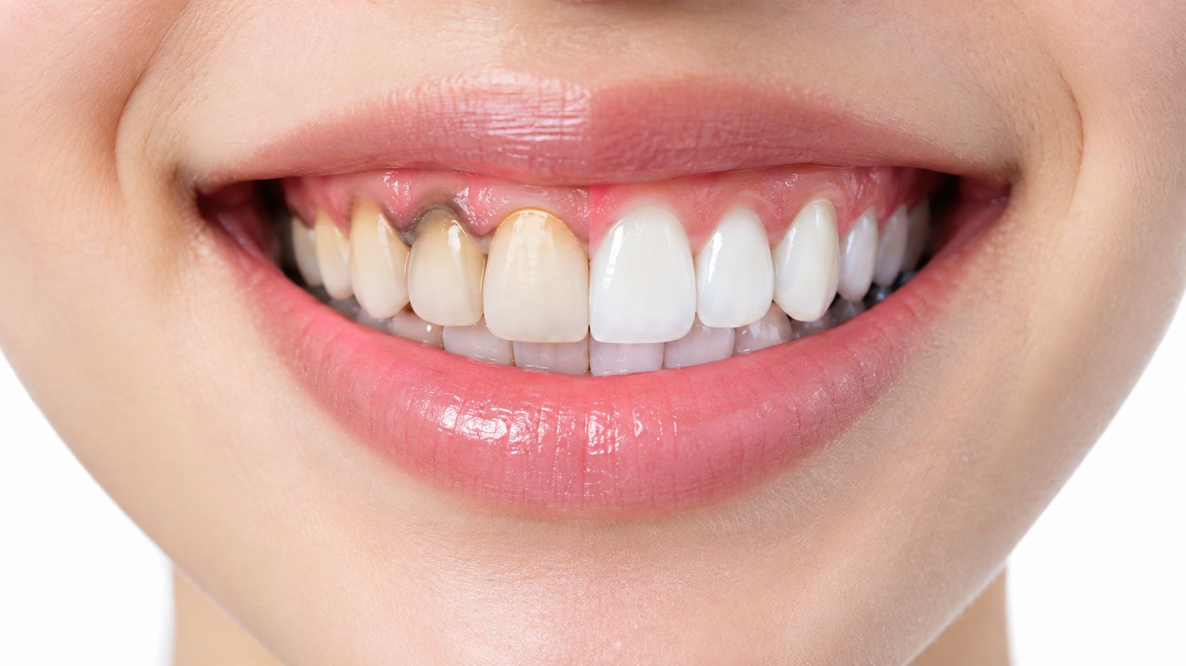
A dark line near the gumline is one of those changes people notice in the mirror and can't quite explain. It may look like a shadow, a stain, or a thin band of something “stuck” where the tooth meets the gum, and that uncertainty is usually what drives the search for a dentist near me in Manhattan or New York.
In a prosthodontic chair, that complaint gets sorted by location first, not by guesswork. A line that sits on the tooth, one that belongs to a crown or filling edge, and one that lives in the gum tissue itself are three different problems, even if they look similar at first glance.
Table of Contents
- When a Dark Line at the Gumline Becomes a Real Concern
- Where the Color Actually Lives and Why That Matters
- Surface Causes on the Tooth Itself
- Restoration Margins and Crown-Related Shadows
- When the Darkness Is in the Gum Tissue
- What a Real Diagnostic Visit Looks Like
- Treatment Options by Origin and When Each Applies
- Next Steps for Patients on the Upper East Side
When a Dark Line at the Gumline Becomes a Real Concern
A patient often notices it while brushing at night or checking a front tooth under bright bathroom light. The reaction is usually the same, a quick worry that the tooth is decaying, the crown is failing, or the smile has changed in a way that can't be reversed.
That concern is reasonable. A dark band at the gumline can be something simple, but it can also point to a problem that needs cleaning, repair, or further evaluation by a prosthodontist or emergency dentist in Manhattan.
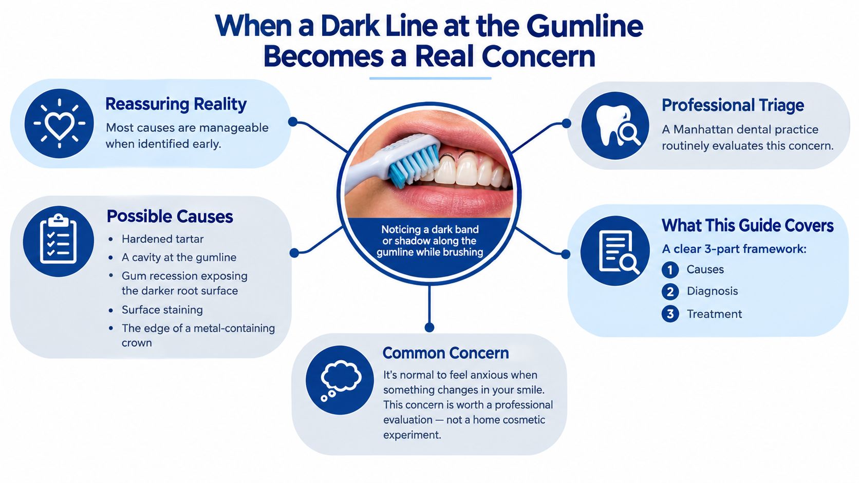
A dark line at the gumline is not automatically decay. Common explanations include hardened tartar, a cavity occurring at the gumline, gum recession that exposes the naturally darker root surface, surface staining, or the edge of a metal-containing crown. Aspen Dental's overview of dark lines near the gumline makes the same practical point, visual similarity alone doesn't tell the whole story.
Practical rule: if the line is changing, rough, or tied to bleeding, sensitivity, swelling, bad taste, or looseness, it deserves a dental exam rather than a whitening kit.
For a patient in the Upper East Side, that distinction matters because the next step isn't the same for everyone. A stain may need polishing, a dark crown margin may need restorative planning, and a gum lesion may need a very different kind of evaluation from a pediatric dentist, endodontist, or restorative team.
Where the Color Actually Lives and Why That Matters
The most useful question is not "Why is this tooth dark?" It's "Where is the dark color sitting?" That answer separates a surface deposit from a restoration edge or a change in the gum tissue, and it narrows the treatment path immediately.
Three locations, three different problems
| Origin location | Visual clue | Most common cause |
|---|---|---|
| Tooth surface | Rough band, plaque-like buildup, or removable stain | Calculus or extrinsic stain |
| Restoration margin | Thin dark line next to a crown or filling | Visible margin, recession, or shadowing |
| Gum tissue | Patch or band in the pink tissue itself | Gingival pigmentation |
That simple map prevents a lot of false alarms. A dark line on the tooth can be cleaned, a dark line at a crown edge may need prosthodontic attention, and a pigmented gum lesion belongs in a different diagnostic bucket altogether.
Why the diagnosis changes with the anatomy
If the color is on the tooth, the dentist looks for plaque, calculus, surface staining, root exposure, or decay. If the color appears to come from a restoration edge, the question becomes fit, margin design, tissue level, and whether the esthetic result can be improved without harming the tooth. If the color is in the gingiva itself, the focus shifts to tissue behavior, symmetry, and whether the lesion is stable or suspicious.
Clinical shortcut: tooth surface problems often respond to cleaning or restorative care, crown-margin problems often need prosthodontic planning, and gum-tissue discoloration may need observation or biopsy.
That is why a Manhattan dental office that handles restorative dentistry, cosmetic dentistry, and gum evaluation under one roof can save time. The complaint may sound cosmetic, but the solution depends on anatomy, not on a whitening assumption.
Surface Causes on the Tooth Itself
When the discoloration sits on the tooth, the first question is whether it can be removed or whether it represents a structural issue. That difference matters because a dark surface can be harmless staining, hardened calculus, or a cavity beginning at the gumline.
Calculus and black stain
Tartar, also called calculus, forms when plaque hardens on the tooth surface. Once mineralized, ordinary brushing and flossing generally can't remove it, and dark deposits near or below the gumline can take on brown or black color when they pick up pigments from coffee, tea, red wine, tobacco, and other dark foods or drinks. Crest's patient guidance on dark or black gums notes that blood and inflammatory fluid can also make the area look darker, which is why a dark band near irritated gums doesn't prove the tooth itself is “dead.”
Black stain is a separate pattern. It's an extrinsic, plaque-associated discoloration that can appear as a thin dark line and may recur after polishing if plaque retention, calculus, bleeding, or irregular restorative margins remain. A professional cleaning can test whether the color lifts off the surface, which is the main practical clue.
Root exposure and gumline decay
A recession line can expose darker root dentin, which naturally looks different from enamel. That can make a healthy tooth look stained even when the underlying issue is loss of gum coverage. Cervical decay is different, because it's a breakdown of the tooth structure near the neck of the tooth and needs restorative treatment, not just cleaning.
What to notice at home
- Removable versus fixed: plaque and stain may reduce with professional care, calculus stays firmly attached.
- Smooth versus rough: a rough band suggests calculus or a defect, not just color.
- Symptoms: sensitivity or a bad taste raises concern for decay or inflammation.
The key point is simple. A dark tooth at the gumline can be cleaned only when the problem is on the surface. If the color is structural, the plan changes.
Restoration Margins and Crown-Related Shadows
A lot of readers focus on staining and miss the most common prosthodontic explanation, the visible edge of an older crown or filling. In the chair, this is the category that most often needs a careful look rather than a cosmetic impulse.
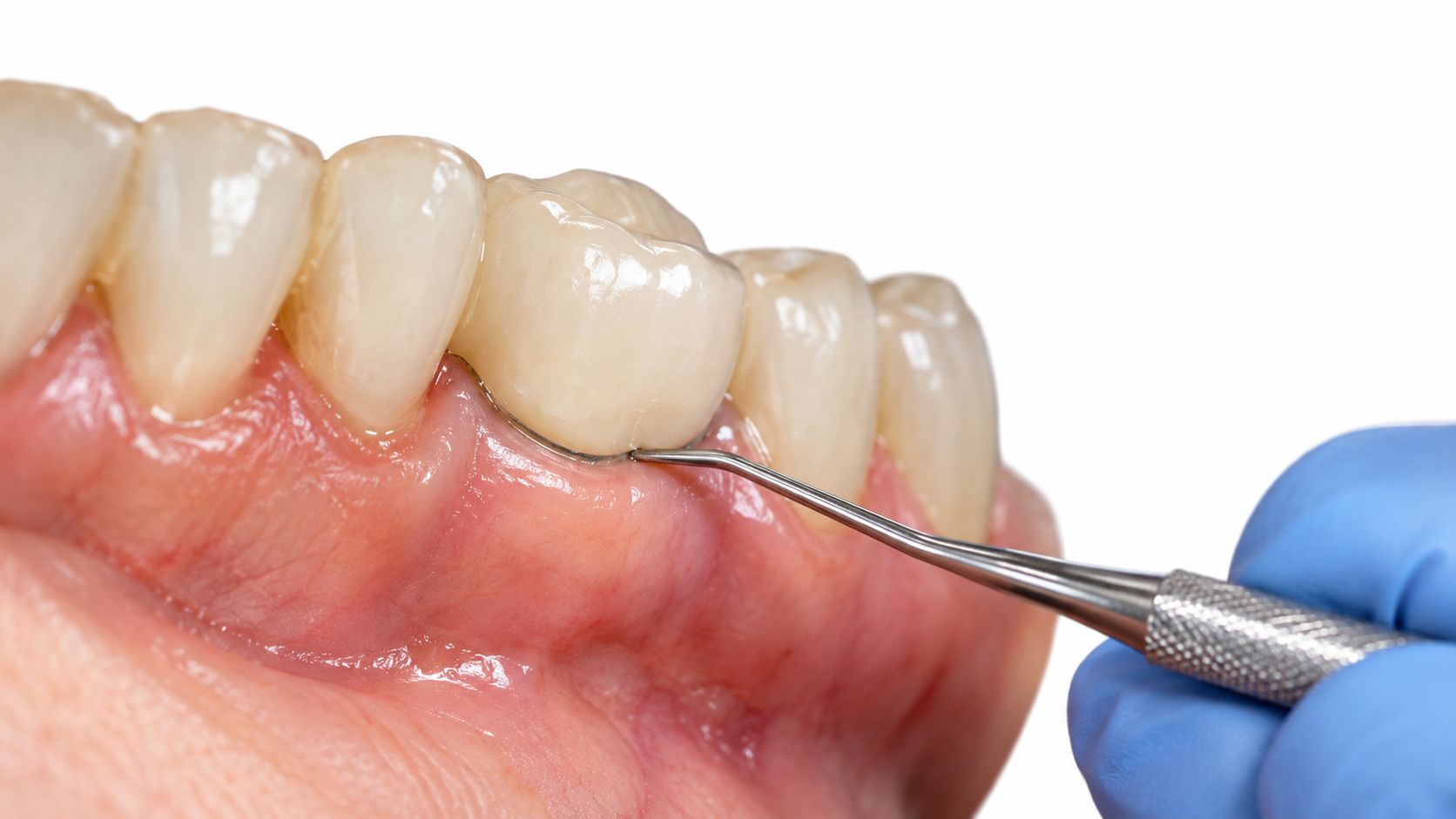
A thin dark line around a crown can reflect a visible metal-ceramic margin, gum recession exposing a darker root or restoration edge, or shadowing through thin gum tissue; whitening generally cannot change the color of a crown margin or exposed restorative material. That restorative distinction is described in a prosthodontic review of crown-edge discoloration, and it's why whitening alone so often disappoints.
Stable line or failing margin
A narrow, stable line without symptoms may be monitored, especially if the restoration still fits well and the tissue is healthy. A widening line with sensitivity, swelling, drainage, bad taste, or recession deserves earlier evaluation because the problem may be marginal leakage, an open edge, or tissue breakdown around the restoration.
What can be done
Sometimes the answer is simple observation. Sometimes a polish or margin refinement helps. In other cases, the crown needs replacement, especially if the esthetic goal is to eliminate a visible dark border or if the restoration no longer supports long-term health.
Prosth & Co.’s crown and bridge care fits that kind of planning, because the question isn't just “Can the color be covered?” It's “Can the tooth, gum, and restoration work together in a way that will hold up?”
Restoration-related darkness is often the place where cosmetic fixes fail. The color may not be in the enamel at all, so bleaching won't touch it. In those cases, the right solution is a prosthodontic one, not a whitening one.
When the Darkness Is in the Gum Tissue
A dark line at the gumline is not always in the tooth or the restoration. When the pigment sits in the gingiva, the question shifts to tissue diagnosis, not whitening or margin correction.
Stable pigmentation versus suspicious change
Physiologic gingival pigmentation is usually symmetric, stable, and otherwise normal in shape and texture. It reflects melanin production and often needs no treatment. A lesion that appears suddenly, changes quickly, or looks irregular deserves a closer look.
Tobacco-associated melanosis has been reported in approximately 20-30% of chronic tobacco users, and focal, asymmetric, irregular, enlarging, newly appearing, bleeding, ulcerated, or multi-colored pigmented lesions require clinical evaluation. The oral pathology review on pigmented lesions also stresses careful documentation and follow-up when the diagnosis is not obvious.
What the clinician looks for
A dentist or specialist usually documents:
- Distribution: one side or both, focal or diffuse.
- Surface features: flat, raised, smooth, irregular, or ulcerated.
- History: onset, tobacco exposure, medication history, and change over time.
- Context: whether teeth, restorations, or gums are involved together.
A new, one-sided, rapidly changing, or symptomatic gum darkening should be evaluated promptly. Home cosmetic treatment is the wrong choice when the pattern does not fit a benign diagnosis.
If the appearance raises concern, biopsy or referral is the safer path. That is how a gum-origin pigment problem gets handled, and it is different from treating stain on enamel or shadow at a crown edge.
What a Real Diagnostic Visit Looks Like
A proper visit starts with examination, not assumptions. The same dark line can be stain, calculus, root exposure, a crown margin, or a gum lesion, so the clinician has to sort location, texture, and tissue health before recommending anything.

The steps that matter
A diagnostic visit typically includes a full exam, periodontal probing, and careful inspection of crown and filling edges. When decay, root involvement, or subgingival calculus is suspected, dental X-rays such as bitewings or periapicals help show what the eye can't see.
A 2020 review of global periodontal-health data reported calculus in 50.3% of adolescents, 44.6% of adults, and 31.9% of older adults in the review of periodontal-health data, which is one reason a dark line below the gumline often turns out to be a professional-cleaning problem rather than a cosmetic one.
What patients usually leave with
The benefit of a real exam is clarity. Instead of a vague “watch it,” patients usually leave with a likely diagnosis, a treatment path, and a sense of whether the issue belongs in preventive care, restorative care, periodontal care, endodontics, or oral surgery.
If digital photos or visual case review are used, the difference can be even clearer because the patient can see what the dentist sees. That kind of documentation is especially useful when a line is subtle, when several restorations are aging at once, or when a cosmetic change needs to be balanced against long-term maintenance.
Treatment Options by Origin and When Each Applies
Treatment should follow cause, not guesswork. A dark line at the gumline can look similar from across the room, but the right fix depends on whether the problem sits on the tooth, at the restoration edge, or in the gum tissue.
Matching the issue to the treatment
| Origin | Typical treatment | Specialist involved |
|---|---|---|
| Tooth surface | Scaling, polishing, hygiene instruction, restorative care if decay is present | Hygienist, general dentist, periodontist |
| Restoration margin | Monitoring, margin refinement, crown replacement, or full-mouth rehabilitation | Prosthodontist, restorative dentist |
| Gum tissue | Observation, tobacco-cessation support, depigmentation, biopsy or referral if suspicious | Dentist, periodontist, oral surgeon |
For surface deposits, scaling and polishing are the starting point. If decay is present at the gumline, the treatment may be a filling, crown work, or, when the tooth can't be saved, tooth extraction followed by replacement planning such as a dental implant or bridge.
For restoration-related lines, the decision is more nuanced. A stable margin may be observed, but a failing edge, poor crown fit, or aging restorative work can justify repair or replacement. That's where a prosthodontist or cosmetic dentist near me search often leads patients to a better long-term answer than bleaching ever could.
For gum pigmentation, observation is appropriate when the tissue looks physiologic. Tobacco-related pigmentation should prompt tobacco-cessation support, and suspicious lesions need biopsy or specialist evaluation. If the appearance sits at the junction of teeth and gums, coordinated care matters most, because periodontal, restorative, and surgical concerns can overlap.
Prosth & Co. is one Manhattan example of that model, with restorative, prosthodontic, periodontic, endodontic, pediatric, and oral surgery services under one roof. That kind of team structure helps when a dark line turns out not to be one problem, but several small ones at once.
Next Steps for Patients on the Upper East Side
The simplest takeaway is this. A dark line at the gumline is usually explainable, but the cause has to be identified before anyone talks seriously about whitening. A professional exam is the only reliable way to separate stain from calculus from decay from a crown margin or gum pigmentation.
Before a first visit, bring a list of restorations, any symptoms and when they started, photos if the line has changed, and your insurance and history forms. If the tooth is sensitive, the gum is bleeding, or the line seems to be widening, don't wait for it to “go away” on its own.
Prosth & Co. on Manhattan's Upper East Side, at 47 E 77th St #207 near the 77th Street 6 train, offers weekday care from 9:00 AM to 6:00 PM with urgent availability. For patients in New York looking for a dentist in Manhattan, an emergency dentist, or a prosthodontist who can sort out a dark line at the gumline with the right mix of cleaning, restoration, and diagnosis, that local access matters.
If a dark line near your gumline has you second-guessing what's stain, what's tartar, and what might need repair, Prosth & Co. can evaluate the area and match the treatment to the actual cause. Schedule a visit through Prosth & Co. for a clear exam, practical next steps, and care that respects both health and esthetics.

Founder and lead prosthodontist of Prosth & Co. on Manhattan's Upper East Side. DDS, Columbia University; Prosthodontics certificate and MMSc in Oral Biology, Harvard School of Dental Medicine; Implantology fellowship, Columbia University.
Meet Victoria →