A routine cleaning can change fast when the gums do not behave like routine gums. A patient comes in from the Upper East Side thinking the issue is just tenderness around a back tooth, then the chart shows a deep pocket, bleeding on probing, and an x-ray pattern that suggests the bone support is no longer flat and stable. At that point, the conversation shifts from cleaning alone to whether the area needs osseous surgery to make it maintainable again.
That conversation matters because deep pockets are not just a number on a periodontal chart. They're a sign that plaque and inflammation are living in a space the patient can't clean well enough at home, and that's where treatment planning gets more specific. For patients comparing options in Manhattan, a helpful starting point is also understanding gum recession causes and prevention, since recession and pocketing often travel together in the same diseased area.
Table of Contents
- When a Deep Pocket Changes the Plan
- What Osseous Surgery Actually Does
- How the Procedure Works Step by Step
- Osseous Surgery vs Non-Surgical and Regenerative Options
- Expected Results and Trade-Offs
- Risks, Recovery, and Aftercare
- Candidacy and Booking a Consultation in Manhattan
When a Deep Pocket Changes the Plan
A patient can feel like everything is fine, then the hygienist measures a pocket around a lower molar and the picture changes quickly. Bleeding on probing, a persistent pocket after earlier care, and horizontal bone loss on a periapical film usually mean the area needs more than another round of cleaning. In that setting, the question becomes whether the bone shape itself is keeping the gum from settling into a healthy, manageable contour.
That is where osseous surgery enters the discussion. In plain terms, the surgeon reshapes the supporting bone so the gum can re-adapt at a shallower depth, with contours that are easier to keep clean. The goal is not to “remove infection” in a vague sense, it is to change the anatomy that lets the pocket persist.
Practical rule: if the pocket stays deep after non-surgical therapy, the defect shape matters more than the label on the chart.
For a Manhattan patient, that usually means the decision is made after a period of scaling and root planing, not instead of it. The visit is about asking whether the tissue has enough support to heal into a shape that a toothbrush, floss, and maintenance visits can control. When the answer is no, bone recontouring becomes the next sensible step.
What Osseous Surgery Actually Does
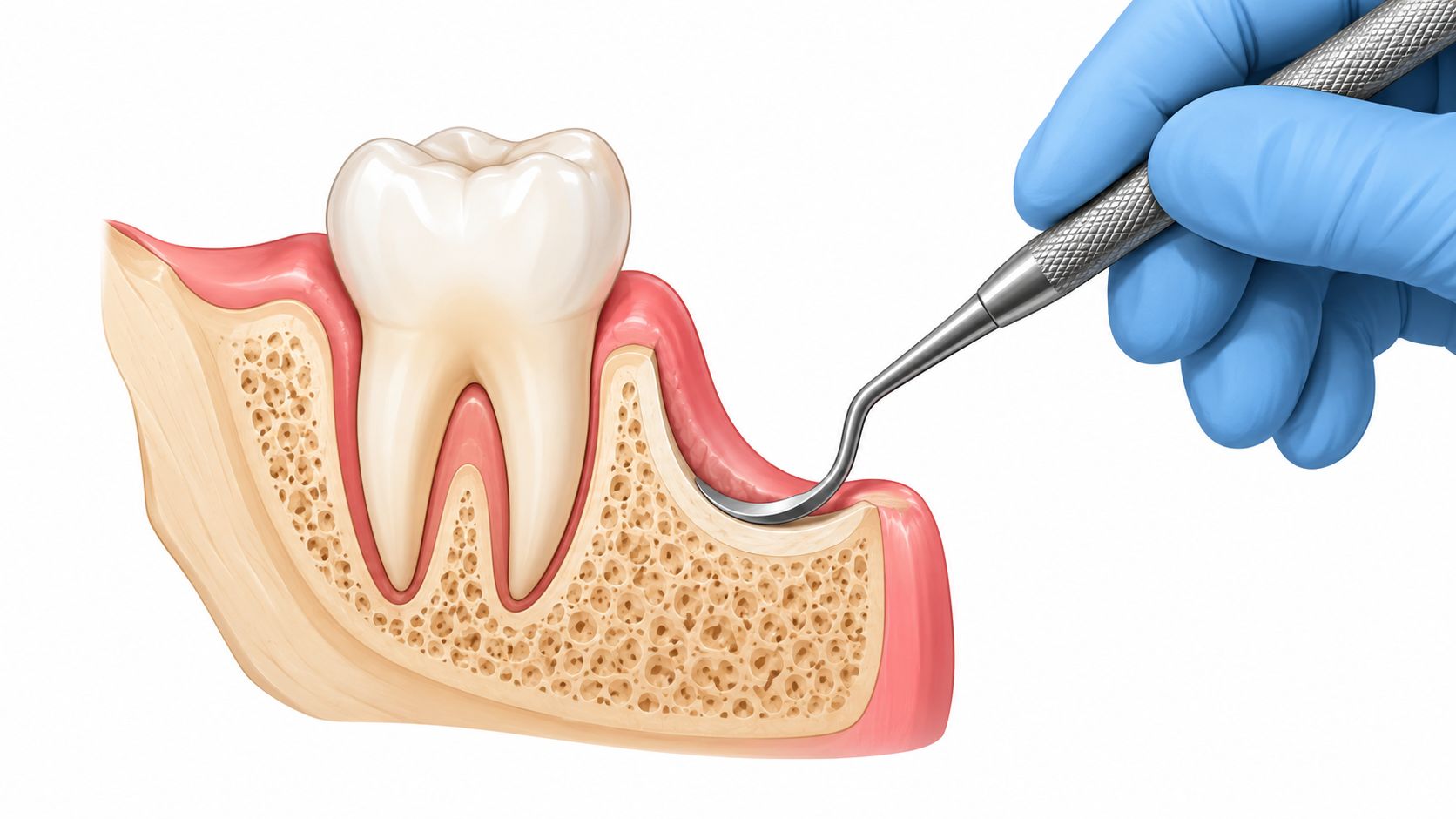
Osseous surgery is a resective periodontal procedure. It combines osteoplasty, which reshapes bone without taking away supporting height, and ostectomy, which removes small amounts of crestal bone when needed so the final contour is more physiologic and easier to maintain. Think of it less like rebuilding from scratch and more like leveling a foundation so the siding can sit flat.
That distinction matters because the aim is pocket reduction, not regeneration of lost bone. The tissue is being asked to heal over a bone outline that matches the gum line better, so plaque control is more realistic after surgery. Patients who still have residual pockets after scaling and root planing are the classic candidates, especially when the defect shape makes daily cleaning difficult.
How the anatomy changes
The bony work is done with a clear endpoint in mind, a contour that supports a healthier gingival margin after healing. That usually means the gum edge sits a little farther down the tooth than before, which is why teeth can look longer afterward. Root sensitivity can follow, especially when areas that were previously covered become more exposed.
The trade-off is honest and direct. A shallower pocket is easier to keep clean, but the visible gumline may sit lower than it did before surgery. In the right case, that trade is worthwhile because maintenance becomes realistic instead of aspirational.
The procedure is also guided by established periodontal technique. Standard descriptions place it as a full-thickness flap procedure with ostectomy and osteoplasty, which is why the surgeon has to anticipate the final contour before the tissue is put back in place. That's the reason flap design and final suturing matter so much, they're part of the architecture, not an afterthought.
How the Procedure Works Step by Step
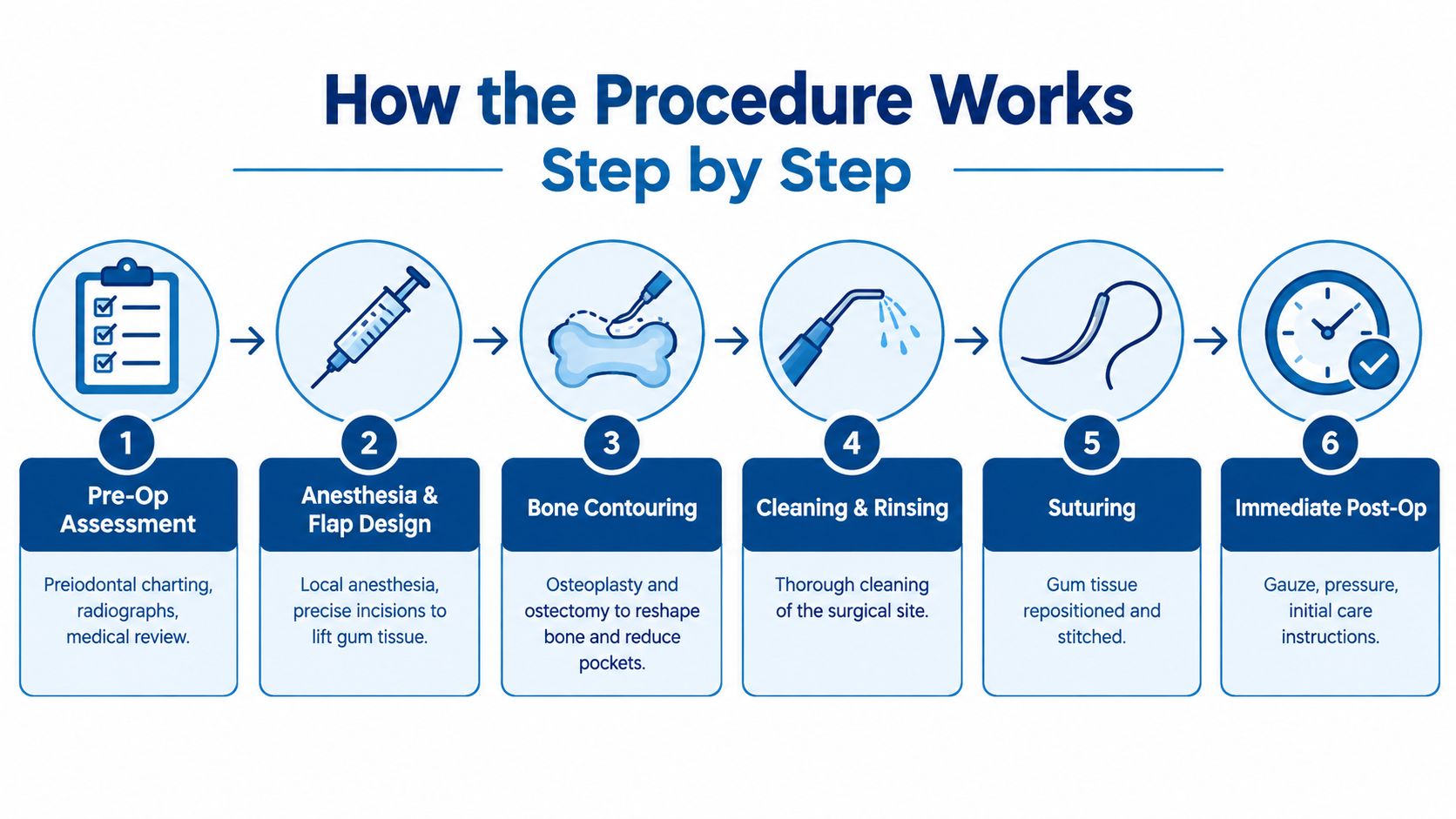
The appointment starts with periodontal charting, radiographs, and a medical review. That first pass tells the team which teeth have isolated defects, which ones have generalized bone loss, and whether the area looks better suited to resection or another pathway. For a patient, this is the planning stage that prevents guesswork once the flap is open.
After numbing, the surgeon makes precise incisions to lift the gum tissue and expose the bone. A sulcular or modified papilla preservation incision may be used depending on the site, because access has to be good enough to correct the defect without tearing the tissue or compromising closure. Under anesthesia, patients usually feel pressure rather than sharp pain.
What happens once the site is open
The next step is debridement. Granulation tissue is removed, and the root surfaces are cleaned so the site is free of inflamed tissue before the bone is reshaped. That matters because bone correction without a clean surgical field doesn't solve the problem that created the pocket in the first place.
Bone contouring follows with hand and rotary instruments, plus continuous irrigation. The surgeon flattens ledges, smooths irregularities, and removes crestal bone where needed to create a contour that will support a maintainable gumline. Depending on the site, a periodontal dressing may be placed, but sutures do the heavy lifting in holding the tissue in its new position.
Healing depends as much on the closure as on the contour. If the flap is not positioned to match the new bone shape, the pocket can come back as the tissue settles.
Most quadrant appointments run about 1 to 2 hours, though the actual time depends on how many teeth are involved and how much reshaping is needed. The patient's role is simple, stay still, stay numb, and follow the closure instructions carefully so the tissue can heal in the intended position.
Osseous Surgery vs Non-Surgical and Regenerative Options
Not every deep pocket should be treated the same way. Some defects respond to non-surgical care, some are better suited to regeneration, and some are most predictable with resective contouring. The right choice depends on morphology, not on a one-size-fits-all “deep cleaning surgery” label.
| Option | Best For | Esthetic Impact | Key Trade-Off |
|---|---|---|---|
| Scaling and root planing | Early disease, shallower pockets, areas that may settle with meticulous maintenance | Usually preserves the gumline better | May not correct anatomy that keeps a pocket open |
| Osseous surgery | Residual pockets, shallow craters, areas where reshaping makes hygiene possible | Can create recession and longer-looking teeth | Gains access by sacrificing some tissue height |
| Regenerative approaches | Contained vertical defects with walls that can support new fill | Better at preserving architecture in selected sites | Not every defect has the anatomy regeneration needs |
| Fiber-retention or conservative flap approaches | Situations where tissue preservation is a priority | Often more favorable for esthetics | May not reduce the pocket as aggressively |
For patients comparing periodontal and restorative planning, an interdisciplinary team may also review whether the site needs bone augmentation for future treatment. The bone grafting approach used in restorative planning is a different tool from osseous resection, but the same principle applies, anatomy drives the choice.
A newer layer of nuance is that furcation involvement alone doesn't automatically mean resective surgery is the answer. Recent review work has pointed out that defect type, long-term tooth retention, and patient tolerance for recession all need to be weighed together. That's especially relevant in molars, where a “surgery or no surgery” framing misses the core decision.
Expected Results and Trade-Offs
The outcome patients usually feel is simpler access to cleaning. Once healing is complete, osseous surgery can reduce pocket depth and reshape areas that trap plaque. Classic periodontal reporting summarized mean reductions of 2.4 ± 0.12 mm on one side and 2.05 ± 0.11 mm on the other in a long-term study, with very small amounts of vertical bone loss after remodeling, as reported in the periodontal literature Pocket Dentistry. Those numbers matter because they reflect the main goal of resective treatment, better access with limited sacrifice of supporting anatomy.
The trade-off is usually visible. A flatter gumline can leave teeth looking longer, and root exposure can make cold sensitivity more noticeable. In the Manhattan office, that conversation is part of the plan before surgery, especially in the front of the mouth where recession is easier to see and harder to ignore. That change is not automatically a complication, it is often the cost of making a defect maintainable.
What patients usually notice
- Cleaner access: brushing and flossing reach the area more easily.
- Less trapped inflammation: the site can settle into a more stable contour.
- More maintenance: the area still needs careful home care and regular professional follow-up.
- Possible esthetic change: black triangles or visible root surface can show, especially in front teeth.
The maintenance burden is real. A reshaped contour stays healthy only if plaque control stays consistent, and periodontal maintenance visits stay part of the long-term plan. For patients who want practical reminders about what that routine looks like, periodontal maintenance guidance is a useful reference.
Risks, Recovery, and Aftercare
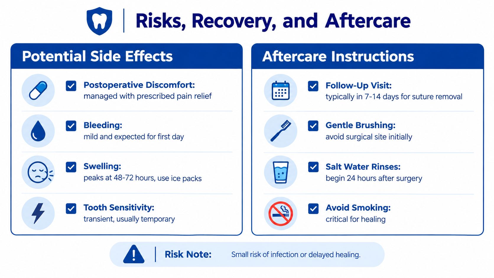
Recovery is usually manageable, but it still counts as surgery. Expect some soreness, mild bleeding, swelling, and short-term sensitivity, especially if the procedure leaves root surfaces a bit more exposed as the tissue settles. Infection and delayed healing are uncommon, but they can happen. New swelling, pain that worsens instead of easing, or bleeding that does not stop should prompt a call to the office.
The first day is about rest, ice, and taking medication exactly as directed. The next few days usually call for soft foods and a lighter chewing load while the area remains tender. Sutures are often removed in about one to two weeks, and brushing and flossing return in stages, not all at once.
A practical aftercare rhythm
- Pain control: use the prescribed or recommended regimen exactly as directed.
- Oral hygiene: keep the rest of the mouth clean while protecting the surgical site early on.
- Salt rinses: begin only when instructed, usually after the first day.
- Smoking avoidance: do not smoke during healing, since it interferes with tissue repair.
- Follow-up: keep the maintenance visits so the new contour does not collect plaque.
A steady home routine matters as much as the surgery itself. Patients who want a simple reminder for what to keep doing between visits can use support your smile between cleanings as a practical reference.
Candidacy and Booking a Consultation in Manhattan
The strongest candidates are patients with persistent pockets above 5 mm after scaling and root planing, localized angular bone loss that can be reshaped, and a home-care routine that's already fairly reliable. The best outcomes usually happen when the defect is anatomically suitable and the patient is willing to accept some recession in exchange for long-term access. Smokers who are unwilling to change habits, patients with uncontrolled diabetes, and cases with widespread horizontal defects often need a different plan.
At Prosth & Co. on the Upper East Side, the evaluation starts with full periodontal charting, including six-point probing, plus periapical and panoramic imaging to map the defect. The team also talks through esthetic priorities, especially how much recession the patient can tolerate in the visible smile, because that changes whether resective or regenerative care makes more sense. If future restorations are planned, the prosthodontic team can review the site with the periodontal findings so the final result supports both health and appearance.
Cost conversations should be direct. Periodontal surgical codes are sometimes partially covered by insurance, but patients often still have an out-of-pocket portion, especially when the case involves detailed imaging or multiple teeth. That's normal, and it's better to know it before treatment than after.
Bring previous cleaning records, x-rays, and any notes from a periodontist or general dentist if they're available. The first visit usually includes records review, probing, and discussion, not surgery the same day unless the clinical situation has already been fully worked up. For patients in Manhattan trying to choose the next step with confidence, that first consultation is the right place to sort out whether osseous surgery, regeneration, or a more conservative approach will best serve the tooth.
Prosth & Co. provides periodontal evaluation, restorative planning, and coordinated surgical care on Manhattan's Upper East Side, with treatment decisions built around health, function, esthetics, and long-term maintenance. If a deep pocket, recession, or difficult molar defect is part of the problem, Prosth & Co. can review the site, explain the trade-offs clearly, and help the patient decide on the most appropriate next step.

Founder and lead prosthodontist of Prosth & Co. on Manhattan's Upper East Side. DDS, Columbia University; Prosthodontics certificate and MMSc in Oral Biology, Harvard School of Dental Medicine; Implantology fellowship, Columbia University.
Meet Victoria →